 Medicine, Psychology and Physiology
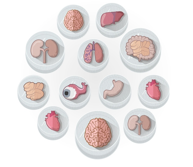
Illustrations by Claire Welsh/Nature
It was an otherwise normal day in November when Madeline Lancaster realized that she had accidentally grown a brain. For weeks, she had been trying to get human embryonic stem cells to form neural rosettes, clusters of cells that can become many different types of neuron. But for some reason her cells refused to stick to the bottom of the culture plate. Instead they floated, forming strange, milky-looking spheres.
“I didn't really know what they were,” says Lancaster, who was then a postdoc at the Institute of Molecular Biotechnology in Vienna. That day in 2011, however, she spotted an odd dot of pigment in one of her spheres. Looking under the microscope, she realized that it was the dark cells of a developing retina, an outgrowth of the developing brain. And when she sliced one of the balls open, she could pick out a variety of neurons. Lancaster realized that the cells had assembled themselves into something unmistakably like an embryonic brain, and she went straight to her adviser, stem-cell biologist Jürgen Knoblich, with the news. “I've got something amazing,” she told him. “You've got to see it.”
Lancaster and her colleagues were not the first to grow a brain in a dish. In 2008, researchers in Japan reported1 that they had prompted embryonic stem cells from mice and humans to form layered balls reminiscent of a cerebral cortex. Since then, efforts to grow stem cells into rudimentary organs have taken off. Using carefully timed chemical cues, researchers around the world have produced three-dimensional structures that resemble tissue from the eye, gut, liver, kidney, pancreas, prostate, lung, stomach and breast. These bits of tissue, called organoids because they mimic some of the structure and function of real organs, are furthering knowledge of human development, serving as disease models and drug-screening platforms, and might eventually be used to rescue damaged organs (see ‘The organoid bank’). “It's probably the most significant development in the stem-cell field in the last five or six years,” says Austin Smith, director of the Wellcome Trust/MRC Stem Cell Institute at the University of Cambridge, UK.
The current crop of organoids isn't perfect. Some lack key cell types; others imitate only the earliest stages of organ development or vary from batch to batch. So researchers are toiling to refine their organoids — to make them more complex, more mature and more reproducible. Still, biologists have been amazed at how little encouragement cells need to self-assemble into elaborate structures. “It doesn't require any super-sophisticated bioengineering,” says Knoblich. “We just let the cells do what they want to do, and they make a brain.”
Growing a gut
This shouldn't come as a major surprise, says molecular biologist Melissa Little at the University of Queensland, Australia. “The embryo itself is incredibly able to self-organize; it doesn't need a template or a map.” That has been known since the early 1900s, when embryologists showed that sponges that had been broken up into single cells could reassemble themselves. But such work fell out of fashion, and modern biologists have focused their attention on purifying cells and growing them in culture — often in flat layers that do little to mimic normal human tissue.
Studying these cells to understand how an organ functions is like studying a pile of bricks to understand the function of a house, says Mina Bissell, a cancer researcher at the Lawrence Berkeley National Laboratory in California. “We should just begin to make the house,” she says. Bissell's work on cultures of breast cells helped to propagate the idea that cells behave differently in 3D cultures than in conventional flat ones. By the mid-2000s, the idea was catching on. The burst of enthusiasm was fuelled by Yoshiki Sasai, a stem-cell biologist at the RIKEN Center for Developmental Biology in Kobe, Japan, who turned heads when he grew a cerebral cortex1, followed by a rudimentary optic cup2 and pituitary gland3 (see Nature 488, 444–446; 2012).
Just a year after Sasai announced his layered cortex, Hans Clevers, a stem-cell researcher at the Hubrecht Institute in Utrecht, the Netherlands, reported the creation of a mini-gut4. The breakthrough stemmed from a discovery in 2007, when Clevers and his colleagues had identified intestinal stem cells in mice. In the body, these cells seemed to have an unlimited capacity to divide and replenish the intestinal lining, and one of Clevers' postdocs, Toshiro Sato, was tasked with culturing them in the lab.
Rather than growing the cells flat, the pair decided to embed them in matrigel, a soft jelly that resembles the extracellular matrix, the mesh of molecules that surrounds cells. “We were just trying things,” Clevers says. “We hoped that we would make maybe a sphere or a blob of cells.” Several months later, when Clevers put his eye to Sato's microscope, he saw more than blobs. The cells had divided, differentiated into multiple types, and formed hollow spheres that were dotted with knobby protrusions. Inside, the team found structures that resembled the intestine's nutrient-absorbing villi as well as the deep valleys between them called crypts. “The structures, to our total astonishment, looked like real guts,” Clevers says. “They were beautiful.”
The mini-guts, reported in 2009, may prove to be a powerful tool in personalized medicine. Clevers and his team are using them to study the effectiveness of drugs in people with cystic fibrosis, who have genetic defects that affect ion channels and disrupt the movement of water in and out of the cells lining the lungs and intestine. The researchers take rectal biopsies from people with the disease, use the cells to create personalized gut organoids and then apply a potential drug. If the treatment opens the ion channels, then water can flow inwards and the gut organoids swell up. “It's a black-and-white assay,” Clevers says, one that could prove quicker and cheaper than trying drugs in people to see whether they work.
He has already used the system to assess whether a drug called Kalydeco (ivacaftor), and 5 other cystic-fibrosis drugs, will work in about 100 patients; at least 2 of them are now taking Kalydeco as a result.
Organoids may also help physicians to choose the best therapies for people with cancer. Earlier this year, Clevers revealed that he had grown a bank of organoids from cells extracted from colorectal tumours5, and David Tuveson, a cancer researcher at Cold Spring Harbor Laboratory in New York, worked with Clevers to generate pancreas organoids using biopsies taken from people with pancreatic cancer6. In both cases, the organoids could be used to find drugs that work best on particular tumours. “What patients are looking for is a logical approach to their cancer,” Tuveson says. “I'm very excited about what we're learning.”
T
he organoid bank
| Organoid | Potential application |
|---|---|
| Cerebral cortex | Understand brain development as well as neurodegenerative diseases and other disorders |
| Intestine | Personalized organoids for identifying patient-tailored drugs |
| Optic cup | Source of retinal tissue for eye therapies |
| Pituitary gland | Source of therapeutic cells for endocrine disorders |
| Kidney | Toxicity testing and a source of tissue for transplantation |
| Liver | Repair of damaged liver |
| Pancreas | Treat diabetes and identify drugs for pancreatic cancer |
| Neural tube | Study nerve development and a source of cell therapies |
| Stomach | Understand stomach development and model gastric disorders such as ulcers |
| Prostate | Predict effective drug combinations for prostate cancer |
| Breast | Understand tumour development |
| Heart | Study cardiac development and how drugs affect it |
| Lung | Model for lung development, maturation and disease |
The small-scale stomach
That excitement is shared by developmental biologist James Wells, who last year reported that he and his team had created an organoid that resembled part of a human stomach7.
Wells started with a different raw material to Clevers, whose organoids arise from adult stem cells that can generate only a limited number of cell types. Wells, who is at the Cincinnati Children's Hospital Medical Center in Ohio, and his colleagues craft organoids from embryonic stem cells, which have the ability to become almost any type of cell. As a result, they have been able to create mini-organs that are more complex.
A decade ago, Wells and his colleagues began trying to coax human embryonic stem cells to form intestinal cells. When the team manipulated two key signalling pathways, the layer of cells produced tiny round buds. Wells noticed that these 'spheroids' mimicked sections of the primitive gut tube, which forms four weeks after conception. This was thrilling, because he realized that he now had a starting point from which to develop a variety of organoids. “Every organ from your mouth down to your anus — oesophagus, lungs, trachea, stomach, pancreas, liver, intestine, bladder — all of them come from this very primitive tube,” he says.
Wells and his colleagues mined the literature and their own experience to determine what chemical cues might send these gut tubes down the developmental path toward a specific organ. Using this strategy, in 2011 the team developed its first human organoid8, an intestine about the size of a sesame seed. But growing a stomach was a bigger challenge. In humans, the organ has two key areas: the fundus at the top, which churns out acid, and the antrum towards the base, which produces many key digestive hormones — and the signalling pathways that lead to one versus the other were unknown. What is more, “the human stomach is different from the stomachs of most animals that we use in the lab”, so there is no good animal model, says Kyle McCracken, a former graduate student of Wells and now a medical student at the centre.
The researchers went for a trial-and-error approach: they made some educated guesses and painstakingly tested different combinations of growth factors. Eventually, the effort paid off. In a 2014 paper7, Wells and his team revealed that they had created organoids that resembled the antrum. Using these as a model system, the team says that it has figured out the chemical trigger that prompts the development of a fundus. Now the researchers are working to answer other basic questions about stomach development and physiology, such as which factors regulate acid secretion, and they are trying to generate other mini-organs from their primitive gut tubes.
This newfound ability to examine human development excites Daniel St Johnston, a developmental geneticist at the University of Cambridge's Gurdon Institute. “You can actually watch how the cells organize themselves to make complicated structures,” he says — something that is impossible in a human embryo. But most organoids are still single tissues, which limits what developmental biologists can learn, he says. “There are certain questions you can't really address because they depend upon the physiology of the whole organism.”
The baby kidney
Melissa Little has spent more than a decade marvelling at the complexity of the kidney. “It has, in an adult, probably 25–30 different cell types, each doing different jobs,” she says. Tubular structures called nephrons filter fluid from the blood and produce urine. The surrounding space, called the interstitium, holds an intricate network of blood vessels and the plumbing that carries urine away.
In 2010, Little and her colleagues started trying to turn embryonic stem cells into a progenitor cell that gives rise to nephrons. For three years, they tried various combinations and timings of growth factors. “It really took a lot of mucking around to make progress,” she says. But finally, in 2013, the team landed on just the right mixture. Little had been aiming to produce just the progenitor cells. But when she looked in the dish she saw two cell types spontaneously patterning themselves as they would in an embryo. “There was a moment of, 'Oh wow. Isn't that amazing',” she says.
This organoid resembles an embryonic kidney rather than an adult one: it has a mix of nephron progenitors and the cells that give rise to urine-collecting ducts9. “If you want to get them to mature further, that's where the challenge really lies,” Little says. So her team has been working to grow a more-sophisticated version — with blood vessels and interstitium. The hope then is to transplant the mini-organs into mice to see if they will mature and produce urine. “I'm pretty excited about what we can build,” Little says.
Because the kidney plays a key part in drug metabolism and excretion, Little thinks that her mini-kidneys could be useful for testing drug candidates for toxicity before they reach clinical trials. And researchers say that other human organoids, such as heart and liver, could similarly be used to screen drug candidates for toxic effects — offering a better read-out on the response of an organ than is possible with standard tissue culture or animal testing.
But Michael Shen, a stem-cell researcher at Columbia University in New York who has created a prostate organoid, is sceptical that these model systems could completely replace lab animals. Animals can show how a therapy affects the immune system, for example, something that organoid systems cannot currently do. “You want to be able to validate your experimental findings in an in vivo system,” he says. “I view that as a rigorous test.”
Little livers
Takanori Takebe was inspired to grow a liver after a chilling spell in New York. While working in the organ-transplantation division at Columbia University in 2010, Takebe saw people die from liver failure owing to a lack of organs. “That was a sad situation,” he says. When he looked into tissue engineering, he thought that the usual methods — seeding cells onto an artificial scaffold — seemed destined to fail. Part of the problem, he says, is that adult liver cells are very difficult to grow. “We cannot maintain it in culture for even a couple of hours.”
Takebe, who took up a research position at Yokohama City University in Japan, decided to work on induced pluripotent stem (iPS) cells, adult cells that have been reprogrammed to behave like embryonic stem cells. He coaxed human iPS cells into forming liver-cell precursors, or hepatoblasts. In the embryo, hepatoblasts rely on a complex symphony of signals from other nearby cells to mature, and Takebe suspected that these support cells would also be necessary to develop a liver in a dish. He and his colleagues mixed hepatoblasts with such cells — called mesenchymal and endothelial cells — and it worked. The team managed to create 'liver buds', structures no bigger than a lentil that resemble the liver of a six-week-old human embryo10. The researchers went on to find that, unlike mature liver cells, such structures can survive in culture for as long as two months.
A liver bud is still a far cry from an entire liver — a hefty, multi-lobed organ composed of tens of billions of hepatocytes. But Takebe hopes that if he can infuse many thousands of buds into a failing organ, he might be able to rescue enough of its function to make a transplant unnecessary. The process seems to work in mice. When Takebe and his group transplanted a dozen of the buds into mouse abdomens, they saw dramatic effects. Within two days, the buds had connected up with the mouse's blood supply, and the cells went on to develop into mature liver cells that were able to make liver-specific proteins and to metabolize drugs. To mimic liver failure, the team wiped out the animals' natural liver function with a toxic drug. After a month, most of the control mice had died, but most of those that received liver bud transplants had survived.
Takebe and his team hope to start human trials in four years. “We will target the children that critically need a liver transplant,” he says. He and his colleagues are currently working to make the liver buds smaller and produce them in huge quantities that they can infuse through the large portal vein that feeds the liver. Takebe thinks that the timeline is “doable”. But Smith says that the process seems rushed, and that the basic biology of these organs needs to be well understood before they are used in the clinic. “It's like running before you can walk,” he says.
Biologists know that their mini-organs are still a crude mimic of their life-sized counterparts. But that gives them something to aim for, says Anthony Atala, director of the Wake Forest Institute for Regenerative Medicine in Winston-Salem, North Carolina. “The long-term goal is that you will be able to replicate more and more of the functionality of a human organ.” Already, the field has brought together developmental biologists, stem-cell biologists and clinical scientists. Now the aim is to build more-elaborate organs — ones that are larger and that integrate more cell types.
And Wells says that even today's rudimentary organoids are facilitating discoveries that would have been difficult to make in an animal model, in which the molecular signals are hard to manipulate. “In a Petri dish it's easy,” he says. “We have chemicals and proteins that we can just dump onto these cells.”